Inhalt
-
Zhang JR, Hardham JM, Barbour AG, and Norris SJ, Antigenic variation in Lyme disease borreliae
by promiscuous recombination of VMP-like sequence cassettes.
-
Zhang JR, Norris SJ , Kinetics
and in vivo induction of genetic variation of vlsE in Borrelia burgdorferi.
-
LymeAlliance.org, LAB TESTS: Reasons Why A Seronegative
Test Result Might Occur.
-
Bauernfeind et al., In vitro activity and stability
against nova beta-lactamases of investigational beta-lactams in comparison
with established compounds.
-
Wiedemann B, Dietz H, Cefotaxime: Unchanged Antibacterial
Activity over Years?
-
Klassifikation der beta-Lactamasen nach Bush
-
Sanders CC, Das veränderliche Bild der mikrobiellen
Resistenz.
-
Brorson O et al., Mattman L, Preac Mursic et al.,
Zystische Formen der Borrelia burgdorferi
-
Riedel M et al., Lyme disease presenting as Tourette's
syndrome.
-
Frieling JTM et al., Differential Induction
of Pro- and Anti-Inflammatory Cytokines in Whole Blood by Bacteria: Effects
of Antibiotic Treatment.
-
Hauser U et al., Validity of interpretation criteria
for standardized Western blots (immunoblots) for serodiagnosis of Lyme
borreliosis based on sera collected throughout Europe.
-
Robertson J; Guy E; Andrews
N; Wilske B; Anda P; Granstrom M; Hauser U; Moosmann Y; Sambri V; Schellekens
J; Stanek G; Gray J, A European Multicenter Study of Immunoblotting
in Serodiagnosis of Lyme Borreliosis.
-
Modai J, Diffusion of 3-quaternary ammonium cephem
antibiotics into cerebrospinal fluid of patients with bacterial meningitis.
-
Wise R, Tissue penetration of the fourth generation
parenteral cephalosporins.
-
Pachner A et al., 2001, Lyme Borreliosis
in Rhesus Macaques: Effects of Corticosteroids on Spirochetal Load and
Isotype Switching of Anti-Borrelia burgdorferi Antibody.
-
Excerpt from Chapter by Alan Steere in: Principles
and Practice of Infectious Diseases, Mandell, Douglas, and Bennett (eds.)
Cell 1997 Apr 18;89(2):275-285
Zhang JR, Hardham JM, Barbour AG, Norris
SJ
(in cache)
Department of Pathology and Laboratory Medicine, University of Texas Medical
School at Houston, 77030, USA.
We have identified and characterized an elaborate
genetic system in the Lyme disease spirochete Borrelia burgdorferi that
promotes extensive antigenic variation of a surface-exposed lipoprotein,
VlsE. A 28 kb linear plasmid of B. burgdorferi B31 (lp28-1) was found to
contain a vmp-like sequence (vls) locus that closely resembles the variable
major protein (vmp) system for antigenic variation of relapsing fever organisms.
Portions of several of the 15 nonexpressed (silent) vls cassette sequences
located upstream of vlsE recombined into the central vlsE cassette region
during infection of C3H/HeN mice, resulting in antigenic variation of the
expressed lipoprotein. This combinatorial variation could potentially produce
millions of antigenic variants in the mammalian host.
Wir haben ein komplexes genetisches System in der Lyme-Borreliose-Spirochäte
Borrelia burgdorferi identifiziert und charakterisiert, das einer umfassenden
Antigenvariation eines Oberflächen-Lipoproteins, VlsE (vls expression
site, vls = vmp-like sequence, vmp
= variable major protein), dient. Wir fanden, daß ein 28 kb ausgedehntes
lineares Plasmid der Borrelia burgdorferi
B31 (lp28-1) eine vmp-ähnliche Sequenzstelle (vls) enthält, das
sehr dem variablen Hauptprotein-System (vmp) für Antigenvariation
des Rückfallfiebers gleicht. Teile von etlichen der 15 nicht-dargestellten
(nicht-ausgeprägten, stummen) vls-Kassetten-Sequenzen oberhalb einer
vls-Ausprägungsposition (Expressionsposition) rekombinierten in die
zentrale vls-Expressionspositions-Kassettengegend während der Infektion
von C3H/HeN-Mäuse, was zur Antigenvariation des dadurch erzeugten
Lipoproteins führte. Diese kombinatorische Variation hätte die
Möglichkeit, Millionen von Antigenvariationen im Säugetier-Wirt
hervorzubringen (Übersetzung des Abstracts: J.G.).
Infect Immun 1998 Aug;66(8):3689-97
Kinetics and in vivo induction of genetic variation of vlsE in Borrelia
burgdorferi.
Zhang JR, Norris SJ
Department of Pathology and Laboratory Medicine and Department of Microbiology
and Molecular Genetics, University of Texas Medical School at Houston,
Houston, Texas 77030, USA.
The Lyme disease agent, Borrelia burgdorferi, is able to persistently
infect humans and animals for months or years in the presence of an active
immune response. It is not known how the organisms survive immune attack
in the mammalian host.
BACKGROUND:
vlsE, a gene localized near one end of linear plasmid lp28-1 and encoding
a surface-exposed lipoprotein in B. burgdorferi B31, was shown recently
to undergo extensive genetic and antigenic variation within 28 days of
initial infection in C3H/HeN mice.
METHODS:
In this study, we examined the kinetics of vlsE sequence variation
in C3H/HeN mice at
-
4 days,
-
7 days,
-
14 days,
-
21 days,
-
28 days,
-
7 months and
-
12 months
postinfection.
RESULTS:
-
Sequence changes were detected by PCR amplification and sequence analysis
as early as 4 days postinfection and accumulated progressively in both
C3H/HeN and CB-17 severe combined immunodeficient (SCID) mice throughout
the course of infection.
-
The sequence changes were consistent with sequential recombination of segments
from multiple silent vls cassette sites into the vlsE expression site.
-
No vlsE sequence changes were detected in organisms cultured in vitro for
up to 84 days.
CONCLUSIONS:
-
These results indicate that vlsE recombination is induced by a factor(s)
present in the mammalian host, independent of adaptive immune responses.
-
The possible inducing conditions appear to be present in various tissue
sites because isolates from multiple tissues showed similar degrees of
sequence variation.
The rate of accumulation of predicted amino acid changes was higher
in the immunologically intact C3H/HeN mice than in SCID mice, a finding
consistent with immune selection of VlsE variants.
http://www.lymealliance.org/Medical/MedCategory4/Med24/med24.html
LAB TESTS: Reasons Why A Seronegative Test Result Might Occur
-
Recent infection before immune response
-
Antibodies are in immune complexes
-
Spirochete encapsulated by host tissue (i.e. lymphocytic celI walls)
-
Spirochete are deep in host tissue
-
Blebs in body fluid, no whole organisms needed for PCR
-
No spirochetes in body fluid on day of test
-
Genetic heterogeneity (300 strains in U.S.)
-
Antigenic variability
-
Surface antigens change with temperature
-
Utilization of host protease instead of microbial protease
-
Spirochete in dormancy phase
-
Recent antibiotic treatment
-
Recent anti-inflammatory treatment
-
Concomitant infection with babesia may cause immunosuppression
-
Other causes of immunosuppression
-
Lab with poor technical capability for Lyme disease
-
Lab tests labeled "for investigational use only"
-
Center for Disease Control criteria is epidemiological, not a diagnostic
criteria
Infection
1991, 19 (Suppl.5): 264-275
In vitro activity and stability against nova beta-lactamases of investigational
beta-lactams in comparison with established compounds
Bauernfeind et al.
Werte sind Minimale Hemmkonzentrationen (MHK) in mg/l
Typ
der
beta-Lactamase |
Cefepim |
Cefpirom |
Cefotaxim |
Cefoxitin |
Piperacillin |
Pip./
Tazobactam |
| TEM-1 |
0.15 |
0.06 |
0.06 |
4 |
>64 |
16 |
| TEM-2 |
0.03 |
0.13 |
0.06 |
4 |
16 |
8 |
| TEM-3 |
2 |
4 |
32 |
16 |
>64 |
8 |
| TEM-4 |
4 |
4 |
32 |
8 |
kein Wert |
kein Wert |
| TEM-5 |
0.5 |
1 |
2 |
16 |
32 |
8 |
| TEM-6 |
4 |
2 |
2 |
4 |
>64 |
16 |
| TEM-7 |
0.5 |
1 |
0.25 |
8 |
>64 |
8 |
|
|
|
|
|
|
|
| SHV-2 |
8 |
16 |
32 |
8 |
>64 |
32 |
| SHV-3 |
2 |
8 |
32 |
16 |
kein Wert |
kein Wert |
| SHV-4 |
2 |
16 |
32 |
4 |
>64 |
64 |
| SHV-5 |
4 |
8 |
32 |
4 |
>64 |
32 |
|
|
|
|
|
|
|
| CMY-1 |
0.25 |
2 |
>64 |
>64 |
>64 |
32 |
|
|
|
|
|
|
|
| CTX-M-1 |
16 |
16 |
>64 |
8 |
>64 |
16 |
Diagn Microbiol Infect Dis 1995; 22:5-12
Elsevier Science Inc, 655 Av of the Americas, New York, NY 10010,
USA
Cefotaxime: Unchanged Antibacterial Activity over Years?
Wiedemann B, Dietz H
Pharmazeutische Mikrobiologie, Universität Bonn, Meckenheimer Allee
168, D - 53115 Bonn, Deutschland
Tab. 1: Auftreten von Extended-Spectrum beta-Lactamasen
| Jahr |
Type |
Klasse |
Repräsentative
Spezies |
Genetischer
Ursprung |
| 1978 |
AmpC |
C |
E. cloacae, Citrobacter
freundii,
Pseudomonas aeruginosa |
Chromosome |
| 1983 |
SHV-2, TEM-3 |
A |
Klebsiella pneumoniae, E.
coli,
C. freundii |
Plasmid |
| 1986 |
K1 |
A |
Klebsiella |
Chromosome |
| 1990 |
AmpC |
C |
K. pneumoniae, E. coli |
Plasmid |
| 1993 |
Metalloenzyme |
B |
P. aeruginosa |
Plasmid |
| 1993 |
OXA-11 |
D |
P. aeruginosa |
Plasmid |
Anmerkung: Klassen A - D nach einer Einteilung, die auf
der Aminosäuren-Sequenz beruht.
Tab. 2: Klassifikation der beta-Lactamasen nach
Bush, 1989
| Bush-Klasse |
Enzym-Typ |
Representative
Enzyme |
| 1 |
Cephalosporinase |
Chromosomal Gram-negativ |
| 2a |
Penicillinase |
Staphylococcus aureus, Pseudomonas
aeruginosa |
| 2b |
Breitband-Spektrum |
TEM-1, -2, SHV-1 |
| 2b' |
Extended Spectrum |
TEM-3, -10, SHV-2, -5 |
| 2c |
Carbenicillinase |
PSE, CARB |
| 2d |
Cloxacillinase |
P. aeruginosa |
| 2e |
Cephalosporinase |
Chromosomal Proteus vulgaris |
| 3 |
Metalloenzyme |
Chromosomal |
| 4 |
Penicillinase |
Chromosomal P. vulgaris |
Continuing Education 1996; E4-X005: 1-8
Bristol-Myers Squibb Company, Route 206 & Providence Line Princeton,
NJ 08543, USA, Tel.: (001) 609 - 252-5141
Das veränderliche Bild der mikrobiellen Resistenz
Sanders, Christie C
Center for Research in Anti-Infectives and Biotechnology, Dept. of Medical
Microbiology and Immunology, Creighton University School of Medicine, Omaha,
Nebraska, USA
...
Die beta-Lactamasen von gram-negativen Bakterien sind vor kurzem in
Übersichtsartikeln beschrieben worden:
-
Sanders CC. beta-lactamases of gram-negative bacteria: new challenges for
new drugs. Clin. Infect. Dis. 1992:14:1089-1099.
-
Sanders CC, Sanders WE Jr. beta-lactam resistance in gram-negative bacteria:
global trends and clinical impact. Clin. Infect. Dis. 1992:15:824-839.
-
Bush K, Jacoby GA, Medeiros AA. A functional classification scheme for
beta-lactamases and its correlation with molecular structure. Minireview,
Antimicrob. Agents Chemother. 1995; 39:1211-1233.
Obwohl sie ziemlich divers sind, schließen die am häufigsten
auftretenden beta-Lactamasen die Enzyme der Bush-Gruppen 1 und 2 ein (siehe
Bush's genanntes Minreview).
-
Bush-Gruppe-1-Enzyme sind die normalerweise chromosomalen
ampC-Enzyme, die nach Induktion vom
Bakterium dargestellt werden
-
in Spezies von Enterobacter,
-
in Spezies von Providencia und
-
in Spezies von Serratia, und
-
in Citrobacter freundii,
-
Morganella morganii und
-
Pseudomonas aeroginosa.
Diese Enzyme sind intrinsisch resistent gegen die beta-Lactamasehemmer
wie Clavulansäure und sind vorwiegend Cephalosporinasen (beta-Lactamasen,
die sich gegen Cephalosporine richten), obwohl sie jede der Hauptklassen-beta-Lactamase-
Antibiotika hydrolysieren.
-
Bush-Gruppe-2-Enzyme sind die am häufigsten angetroffenen Plasmid-kodierten
beta-Lactamasen, TEM-1, TEM-2, SHV-1 und die mutierten Derivate dieser
Enzyme, die extended-spectrum beta-lactamases (ESBL's), die sich aus ihnen
entwickelt haben. Die Bush-Gruppe-2-Enzyme sind intrinsisch empfindlich
gegen beta-Lactamase-Hemmer, obwohl mutierte Formen beschrieben werden,
die unempfindlich gegen die Hemmer sind.
-
Bush-Gruppe-3-Enzyme schliessen die weniger häufig anzutreffenden
metallo-beta-Lactamasen, die für die Resistenz gegen Carbapeneme verantwortlich
sind (siehe Bush's genanntes Minreview).
Es gibt drei Haupt-Mechanismen, durch die Organismen antimikrobiellen Mitteln
widerstehen können (Holmerg SD, Solomon SL, Blake PA. Health and economic
impacts of antimicrobial resistance. Rev. Infect. Dis 1987:9:1065-1078.)
-
Verhinderung der Ansammlung toxischer Konzentrationen der Droge in der
Zelle
-
Veränderungen an den Proteinen der äußeren Membran (nur
gram-negative Bakterien haben eine solche). Beispiel: Imipenem-Resistenz
bei Pseudomonas aeruginosa.
-
Verminderter aktiver Transport der Droge in die Zelle hinein. Beispiel:
Aminoglycosid-Resistenz bei gram-positiven und -negativen Bakterien.
-
Aktiver Transport der Droge aus der Zelle heraus. Beispiel: Tetracyclin
oder Quinilon-Resistenz bei gram-positiven oder -negativen Bakterien.
-
Veränderung des Zielorts der Droge
-
Erwerb eines neuen, weniger empfindlichen Zielorts. Beispiel: Erwerb des
PbP 2a (Penicillin bindendes Protein 2a) in Methicillin-resistenten Staphylococcen.
-
Mutation des Zielorts hin zu einer weniger empfänglichen Form. Beispiel:
veränderte DNA-Gyrase in Quinolon-resistenten gram-positiven und -negativen
Bakterien.
-
Erwerb von vielfachen neuen Genen, die den Zielort verändern. Beispiel:
Vancomycin-Resistenz bei Enterococcen.
-
Produktion von inaktivierenden Enzymen
-
Chloramphenicol-Azetyltransferase,
-
Aminoglycosid-inaktivierende Enzyme,
-
beta-Lactamasen.
....
Resistenz über die Undurchläßigkeit der äußeren
Membrane ist ein wirksamer Resistenz-Mechanismus gegen beta-Lactam-Antibiotika
bei gram-negativen Bakterien, insbeondere in der Kombination mit beta-Lactamasen.
ChemotherapieJournal, 8. Jahrgang, Heft 5, 1999
Abt. für Medizinische
Mikrobiologie und Hygiene, Universitätsklinikum
Ulm, Steinhövelstr. 9, 89075 Ulm
matthias.trautmann@medizin.uni-ulm.de
....
Die essentiellen Penicillin bindenden Proteine (PbP) werden ihrem Molekulargewicht entsprechend als PbP-1, -2 und -3 bezeichnet. Die Blockade nur eines der 3 essentiellen PbP führt nicht notwendigerweise zum Absterben der Bakterienzelle (siehe Abb. 1 in Heinemann M, Trautmann M, 1999)
- Die Antibiotika-Interaktion mit PbP-1 in
einer Konzentration oberhalb der Minimalen Hemmkonzentration (MHK)
führt zu einem relativ schnellen Absterben der Bakterien (Prins et al. 1994). Cefsulodin und
Cephaloridin sind paradigmatisch für
Antibiotika, die primär an PBP-1 binden
und rasch bakterizid wirken.
- Beta-Lactam-Antibiotika, die eine hohe PBP-2-Affinität aufweisen, führen zur Bildung
von rundlichen Zellen, den so genannten
Sphäroplasten ohne einen allzu ausgedehnten Zellwandzerfall. Dadurch bleibt
die Endotoxin-Freisetzung begrenzt (Prins et al. 1994,
Trautmann et al. 1998, Pucci et al. 1991). Carbapeneme wie Imipenem und
Meropenem sind typische Vertreter von
PBP-2-Inhibitoren [50]. Zu den PBP-2-
spezifischen Antibiotika, welche die Bil-
dung von Sphäroplasten induzieren,
gehören außerdem Mecillinam, Clavulansäure und Cefepim, ein Cephalosporin
der vierten Generation (Prins et al. 1994, Pucci et al. 1991).
- Antibiotika mit selektiver PBP-3-Aktivität stören zunächst nur die
bakterielle Septierung und führen daher
zur Bildung von langen, filamentösen
Zellen. Beispiele sind: Aztreonam, Piperacillin, Mezlocillin und bei niedrigeren Antibiotika-
Konzentrationen auch Cefuroxim und die
Cephalosporine der dritten Generation,
Ceftazidim und Cefotaxim Mehrere Arbeitsgruppen konnten zeigen,
dass die Lyse filamentõser, bakterieller
Zellen zu einer schnellen und deutlichen
Zunahme der Endotoxin-Freisetzung
führt, vermutlich aufgrund der großen
Biomasse dieser langen Zellen (Hurley 1992, Trautmann 1998,
Trautmann 1998, Hurley 1993).
Literatur
Hurley JC. Antibiotic-induced release of endo-
toxin: a reappraisal. Clin Infect Dis 1992;15:
840-54.
Hurley JC. Reappraisal of the role of endotoxin
in the sepsis syndrome. Lancet 1993;341:1133-
5.
Prins JM, van Deventer SJ, Kuijper EJ, Speel-
man P Clinical relevance of antibiotic-induced
endotoxin release. Antimicrob Agents Chemo-
ther 1994;38:1211-8.
Pucci MJ, Boice-Sowek J, Kessler RE, Doug-
herty TJ. Comparison of cefepime, cefpirome,
and cefaclidine binding affinities for penicillin-
binding proteins in Escherichia coli K-12 and
Pseudomonas aeruginosa SC8329. Antimicrob
Agents Chemother 1991;35:2312-7.
Trautmann M, Zick R, Rukavina T, Cross AS, et
al. Antibiotic-induced release of endotoxin: in-
vitro comparison of meropenem and other anti-
biotics. J Antimicrob Chemother 1998;41:163-
9.
Trautmann M, Heinemann M, Zick R, Moricke
A, et al. Antibacterial activity of meropenem
against Pseudomonas aeruginosa, including an-
tibiotic-induced morphological changes and
endotoxin-liberating effects. Eur J Clin Micro-
biol Infect Dis 1998;17:754-60.
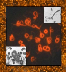
Zystische Formen der Borrelia burgdorferi
Borrelia burgdorferi Cystic Forms
Aussehen - Appearance

Fig. 1: Schema einer Spirochete mit Zyste:
- Querschnitt und
- Draufsicht (unten links)
(nach Preac
Mursic et al 1996 (siehe Two
spherical bodies adhering to the middle of a Borrelia organism),
Brorson,
Brorson 1998 und Atlas
R, Seite 110).
Zeichenerklärung von innen nach außen:
Die Spirochäte Borrelia burgdorferi hat
-
7 - 11 Flagellen (FL) zwischen der ZW und AM, die ihnen eine variable
Spiralform geben. Beim Bewegen der Flagellen verändert sich die Spiralform
analog der einer Schlange bei der Fortbewegung. Bei Umwandlung in die zystische
Form gibt es Stadien, in denen die Flagellen freigelegt, d.h. nicht mehr
von der äußeren Membran eingeschlossen sind (Preac
Mursic et al 1996).
-
ZW und AM sind im periplasmatischen
Raum durch sog. Outer surface Proteine (die Lipoproteine OspA,
OspB, ...) verbunden.
-
Lipopolysaccharide (LPS) sind außen an
der AM angebracht (LPS bei B. burgdorferi).
-
Eine Glycoprotein-Schicht ("S-Layer") umgibt die Borrelie (Grier
T M, Lyme Disease Survival Manual, 1997).
Die Oberflächen außerhalb der Zytoplasmamembran (ZM),
also
- die Zellwand (ZW) und
- die äußere Membran (AM, outer membrane),
werden durch bakterieneigene Lysozyme (auflösende
Enzyme) beim Wachstum aufgelöst. Wenn durch Verwendung von Penicillinen
oder die Wirkung des Immunsystems das Gleichgewicht zwischen bakterieller
Auflösung und Wiederaufbau gestört wird, entstehen zellwand-defizitäre
Formen (L-Formen, auch Spheroplaste
oder Zysten genannt), bei denen die Zytoplasma-Membran (ZM = cytoplasma
membrane) und Flagellen von außen sichtbar werden (siehe auch IDEAS:
THE BACTERIA REVOLUTION, May 28 & June 4, 1999 CBC Radio).
(click here
for English text).
Das Verhältnis von Zysten- und Spirocheten-Volumen variiert in
weitesten Grenzen,
(There is a wide range of relative
cyst-sizes,
Die Zysten können sich von den Spirocheten lösen.
Sie werden dann "Blebs"
genannt.
(Small cysts that have detached from the spirochete
are called "blebs").
Die im Vergleich zu Gram-positiven Bakterien sehr dünne Zellwand setzt
kleinen Molekülen wie den Antibiotika keinen Durchlaßwiderstand
entgegen, während hingegen die äußere und die Zytoplasma-Membran
die Durchläßigkeit sehr aktiv bestimmen.
-
Für beta-Lactam-Antibiotika ist die äußere Membran eine
Barriere.
-
Beta-Lactam-Antibiotika binden sich an die Penicillin-bindenden-Proteine
(PBP) und die Beta-Lactamasen (beta).
-
Die Zielpositionen aller anderen Antibiotika liegen innerhalb der Zytoplasma-Membran.
Bakterien können eine Resistenz gegen diese Drogen dadurch entwickeln,
daß sie eine Ansammlung der Droge im Inneren der Zytoplasma-Membran
verhindern.
Eigenschaften - Properties
of cystic forms
-
Preac Mursic et al
1996
-
Persistors [i.e. B. burgdorferi found after long antibiotic treatments
of the patients] isolated from a great number of patients (60- 80%) after
treatment with antibiotics had an atypical form. The morphological alterations
in culture with penicillin G developed gradually and increased with duration
of incubation.
-
Pleomorphism,
-
the presence of elongated forms and spherical structures,
-
the inability of cells to replicate,
-
the long period of adaptation to growth in MKP-medium and
-
the mycoplasma-like colonies after growth in solid medium (PMR agar)
suggest that B. burgdorferi produce spheroplast-L-form variants.
-
With regard to the polyphase course of Lyme borreliosis, these forms without
cell walls can be a possible reason why
-
Borrelia survive in the organism for a long time and
-
the cell-wall-dependent antibody titers disappear only to emerge again
after reversion.
-
Brorson, Brorson
1998, 1999, 2009
-
Normal, mobile spirochetes were inoculated into spinal fluid. The spirochetes
converted to cysts (spheroplast L-forms) after 1-24 h.
-
When these cystic forms were transferred
to a rich BSK-H medium, the cysts converted back to normal, mobile spirochetes
after incubation for 9 to 17 days, the older the cysts the more time they
needed for reversion. If growth medium is insufficient, no reversion was
observed.
-
Young cysts were observed to be relatively empty except for some spirochete-like
structures, whereas in old cysts these structures will be altered towards
core structures.
-
When cysts are opended forcibly with NaOH, spirochete-like structures will
escape from them.
-
When neuroborreliosis is suspected, it is necessary to realize that B.
burgdorferi can be present in a cystic form, and these cysts have to be
recognized by microscopy. This study may also explain why cultivation of
spinal fluid often is negative with respect to B. burgdorferi.
-
"There is evidence (in vitro) that metronidazol
(Flagyl) will kill the cystic form. This fits with the now well known clinical
observations that Flagyl can be remarkably effective for many chronic Lyme
patients. However, this medication apparently has no effect on intact spirochetes.
Therefore, the trend now is to treat the chronically
infected patient who has resistant disease by combining Flagyl with 1 or
2 other antibiotics to target all forms of Bb. Because there is laboratory
evidence that tetracyclines may inhibit the effect of Flagyl, this class
of medication should not be used in these 2- and 3-drug regimens"
(Burrascano,
1999).
-
-
Alban PS, Johnson
PW, Nelson DR
-
B. burgdorferi cells cannot synthesize fatty acids de novo and serum
is thought to provide a source of fatty acids and lipids.
-
When B. burgdorferi cells were serum-starved (in defined RPMI medium),
-90% of the cells formed spherical cysts within 48 h.
-
Cyst formation was inhibited by tetracycline.
-
Cyst opening and recovery of vegetative cells (cells that are able to divide)
was rapidly induced by the addition of either BSKII medium or rabbit serum.
2.9% to 52.5% of cysts converted into viable cells.
-
Viability was inversely proportional
to cyst age.
-
Data suggest that cells of B. burgdorferi, although possessing a small
genome and extremely limited biosynthetic capabilities, rapidly respond
to conditions of serum starvation by inducing changes in protein synthesis
and cell morphology.
-
Ovcinnikov NM, Delectorskij VV 1968, 1971
-
Formation of protective cysts is widespread in
nature.
-
Can the host harbour cysts of pathogenic
nature?
-
Gruber 1999
-
Theoretically, cystic forms may act as niches
protecting Bb from immediate destruction by antibiotics or immune system.
-
They release spirochetal forms of Bb (by reversion), and the thus newly
developed outer membrane and cell wall components -being TI-1
antigens- can
-
either drive self-organized
feedback oscillations of the immune system or
-
lead to cycles of the elimination
and inflammation imprinted
on the immune system by fluctuations of the endocrine system.
Literatur - Literature
- Cheryl's Lyme Info Web Site, Lyme Files
- Cystic Form File:
These abstracts document the ability of Borrelia burgdorferi (Bb) and related bacteria to"'shape-shift" and change forms in
response to surrounding conditions. These cystic forms shed light on the mechanisms Bb uses to survive antibiotics. The fact that Bb does this gives a
scientific explanation for what many patients experience...the improvement of their medical condition on antibiotics, worsening when off. 17 pages.
-
Pictures of Alternate Forms of Spirochetes:
This file contains photographs of alternate forms of spirochetes --
cystic, granular, bleb forms, etc. Quotations from peer-reviewed studies
are included to aid in understanding how the alternate forms enable
spirochetes to overcome adverse environmental conditions. The ability of
Bb (the Lyme disease bacterium) to transform itself to and from
non-spirochetal forms provides a cogent explanation for relapses after
antibiotic therapy, and for the occurrence of seronegative Lyme disease.
30 pages. Note: This file is fairly large (4 MB)Ê because it contains a
large number of photographs, so be aware that it may take a while to
download.
- Alt Forms Bibliography:
This file is a complete bibliography of over 250 studies (!) with information/observations on alternative forms of
spirochetes. 48 pages
-
Two very extensive literature searches on cystic forms of spirochetes:
I,
II.
-
Cystic Forms: Search Marie Kroun's LymeRICK server
APMIS 2001 May;109(5):383-8
Conversion of Borrelia garinii cystic forms to motile spirochetes in vivo.
Gruntar I, Malovrh T, Murgia R, Cinco M
Institute of Microbiology and Parasitology, Veterinary Faculty,Ljubljana,
Slovenia. gruntaig@mail.vf.uni-lj.si
ABSTRACT - Cystic forms (also called spheroplasts or starvation
forms) andtheir ability to reconvert into normal motile spirochetes have
already been demonstrated in the Borrelia burgdorferi sensu lato complex.
The aim of this study was to determine whether motile B. garinii coulddevelop
from cystic forms, not only in vitro but also in vivo, in cyst-inoculated
mice. The cysts prepared in distilled water were able to reconvert into
normal motile spirochetes at any time during in vitro experiments, lasting
one month, even after freeze-thawing of the cysts. Motile spirochetes were
successfully isolated from 2 out of 15 mice inoculated intraperitoneally
with cystic forms, showing the infectivity of the cysts. The demonstrated
capacity of the cysts to reconvert into motile spirochetes in vivo and
their surprising resistance to adverse environmental conditions should
lead to further studies on the role and function of these forms in Lyme
disease.
From: joanne822@aol.com (Joanne822)
Newsgroups: sci.med.diseases.lyme
Subject: Cysts in T.Pallidum (2 studies)
NNTP-Posting-Host: ladder07.news.aol.com
X-Admin: news@aol.com
Date: 04 Jan 2000 04:14:03 GMT
Organization: AOL http://www.aol.com
Message-ID: <20000103231403.20111.00000314@ng-fv1.aol.com>
Xref: news.gmd.de sci.med.diseases.lyme:63762
British Journal of Venereal Diseases,
1968;44:1-34
Further Study of Ultrathin Section of Treponema Pallidum under the Electron
Microscope
N.M. Ovcinnikov and V.V. Delectorskij
The encystment of treponemes is of immense interest. Under unfavorable
conditions of existence for treponemes (lack of nutrients, the using up
of nutrients for the process of division, after the addition of sera, particularly
immune sera, and small quantities of penicillin, etc.), cysts are formed.
Encystment begins with the treponeme "packing" itself to form a compact
structure (Figs 75, 76) which then becomes covered with a mucous mass (Fig.
74MU). ...
That sharply-marked structural elements of the treponeme and its complex
and characteristic structure indicate that cysts are not a product of degeneration.
In addition, in cultures where there are many cysts, they are very mobile,
which is another argument against degeneration. [as an interpretation of
cysts]
... A treponeme may undergo transverse division in several places along
its length, not merely in one place.
...In actual fact, under unfavourable conditions of existence, treponemes
form real cysts as a method of persistent survival and multiplication,
as occurs not infrequently among protozoa.
..An important proof of the viability of cysts is their motility, as
seen under dark-field and phase-contrast microscopy. When transfers are
made from cultures containing cysts and almost no ordinary spiral forms,
growth of ordinary spiral forms occurs.
...This phenomenon [the formation of protective
cysts] is widespread in nature.
-
The huge majority of cysts in protozoa, according to Dogel (1951), are
a means of protecting their content against unfavourable conditions, but
-
some of them are designed rather to ensure a long period of rest.
-
Later, depending on conditions and when the harmful exposure is past, protective
cysts may become multiplcation cysts.
-
They are not of a purely protective character but also serve for multiplcation...
-
The main factor regulating cyst formation is the exhaustion of food in
the environment.
British Journal of Venereal Diseases, 1971, 47:315-328
Current Concepts of the Morphology and Biology of Treponema pallidum based
on Electron Microscopy
N.M. Ovcinnikov and V.V. Delectorskij
The breakdown into granules [blebs] is especially pronounced under
the action of penicillin and immune sera...
Of extreme importance is the ability of treponemes to form cysts under
unfavourable conditions... Under stressful conditions, the treponeme 'packs'
itself into a compact roll (Fig. 8) and becomes covered with a transparent
mucoid capsule, which resists the penetration of drugs and antibodies.
The organisms may persist in this form for a prolonged period without any
reaction from the host. The encysted treponemes and the host coexist more
or less peacefully, but under propitious circumstances the cysts may be
transformed again into the usual spiral, which damages the cells of the
host and elecits a response.
...Moreover, a cyst may contain laminar bodies of variable dimensions;
we believe that these are stores of nutrients.
...Can the host harbour cysts of
pathogenic treponemes? This is a problem of paramount importance. Even
those authors who admit the possibility of encystment of T. pallidum do
not consider it likely that cysts may exist in host tissues. By means of
electron microscopy we have succeeded in demonstrating the presence of
cysts in a rabbit chancre, and we believe that this observation is of considerable
significance.
The material was taken from an 8-month-old chancre. When examining the
cysts, we could distincly see multi-layered membranes and treponemes cut
in various places. Obviously such a membrane serves as a strong barrier
to drugs.
...In a rabbit chancre T. pallidum may exist both inside and outside
cells.
...The L-forms of T. pallidum do not differ significantly in appearance
from those of other organisms, for example gonococcus, Proteus vulgaris,
etc. When L-forms are transferred to the usual media they soon reverse
to the original forms... Some of them are seen to divide. Thus it is evident
that T. pallidum may produce L-forms under conditions of stress. In our
experiments the organisms were stressed with penicillin, various drugs,
antisera, etc., and the addition of these agents led to the appearance
of L-forms. We have not yet isolated L-forms directly from animals affected
with syphilis, but undoubtedly they can be present in rabbit tissues.
The significance of these data can hardly be overestimated if we recall
that conversion to L-forms alters
-
the clinical course of a disease,
-
the virulence and pathogenicity of the organisms,
-
their antigenic activity,
-
drug resistance,
and other properties, not to mention the diagnostic difficulties presented
by such variants.
The Lancet Vol. 351-February
7, 1998
Lyme disease presenting as Tourette's syndrome
Michael Riedel, Andreas Straube, Markus J Schwartz, Betina Wilske, Norbert
Müller
Lyme borreliosis is often misdiagnosed , both in adults and children. (1)
Central Nervous system manifestations of Lyme disease include neurological
and psychiatric symptoms. (2) Although abnormal movements
have been observed in Lyme disease, (3) a Tourette's
syndrome has not been reported.
A boy at the age of 4 years developed a simple motor tic (blinking)
that resoved within a year without treatment. At the age of 9 years, he
developed
-
multiple orafacial tics including shaking of the head, and several weeks
later a vocal tic occurred.
-
The tics became exacerbated under stress, as typically seen in Tourette's
syndrome.
-
Social disabilities such as loss of impulse control, social withdrawl,
worsened performance at school followed.
He came to hospital at 11 months after onset of symptoms.
-
Serum IgM antibody titres against Borrelia burgdorferi measured by ELISA
were not increased;
-
although IgG antibody titres (ELISA) were increased at 58 U/mL (normal
\< U/ml) and 100 U/mL at another examination 2 weeks later.
-
Immunoflourescence absorption test (IFT) was also increased (1:28 [normal
\<1:16]).
-
IgG immunoblot (4) was positive.
All results indicated an infection with B burgdorferi.
Examination of cerebral fluid
-
showed a slight lymphocytic pleocytosis (16 cells per uL) (5)
which suggested an inflammatory reaction.
-
The CSF: serum IgG ratio for IgG antibodies was 2.0, indicating intraethecal
production of B. burgdorferi specific IgG antibodies, as occurs in neuroborreliosis.
(4)
The boy was treated with intravenous ceftriaxone 2 g daily for 14 days.
-
The tics improved after the sixth dose, and
-
after the tenth dose the tics resolved completely.
-
His social skills returned to normal.
-
Follow-up examinations showed no reccurence of tics or other neurological
or psychiatric disorder.
-
Serum IgG antibody titre tests and IFT tests against B burgdorferi were
11 U/mlL and 1:32 after 1 year.
Rapid efficacy of antibiotic treatment followed by a decrease in Borrelia-specific
antibody titres suggests that the multiple motor and vocal tics were at
least partially caused by the tertiary stage of borreliosis. (5)
Persistence of the tics and increasing severity of the social disabilities
over several months suggest that the first signs of a Tourette-like syndrome
11 months previously were an expression of an early Lyme infection.
Infection with B burgdorferi should be considered in cases of Tourette's
syndrome in endemic areas.
References
-
Shapiro ED, Selzter EG, Lyme disease in children. Semin Neurol 1997;17:39-44.
-
Kaplan RF, Jones-Woodward L,. Lyme encephalopathy; a neuropsychological
perspective. Semin Neurol 1997;17:31-37
-
Fallon BA, Nields JA, Parsons B, Liebowitz MR, Klein DF, Psychiatric
manifestations of Lyme Borreliosis. J Clin Psychiatry 1997;54:263-68
-
Wilske B, Fingerle V, Herzer P, et al. Recombinant immunoblot in
the serodiagnosis of Lyme borreliosis. Med Mikrobiol Immunol 1993; 182:255-70.
-
Pfister HW, Wilske B, Weber K., Lyme borreliosis: basic science
and clinical aspects. Lancet 1994; 363:1031-16
Antimicrobial Agents and
Chemotherapy, July 1997, p. 1439-1443
J. T. M. Frieling, J. A. Mulder, T. Hendrics, J. H. A. J. Curfs, C. J.
van der Linden, R. W. Sauerwein
....
LPS wird von lebenden und wachsenden Bakterien abgegeben, aber ein Abtöten
der Bakterien durch antimikrobielle Medikamente kann zusätzlich LPS
freisetzen. Dies kann zu einem anfänglich schädigenden Einfluß
der antibiotischen Behandlung auf den Zustand des Patienten führen.
Solche Phänomene wurden bei Typhus (1) und vermuteter
Ansteckung durch gram-negative Bakterien beschrieben (2)
und werden unterstützt durch Studien, die erhöhte Konzentrationen
zeigen nach Antibiotika-Behandlung von
-
klinischen (3) oder experimentellen (4
- 7) Infektionen und
-
in in-vitro Modellen (8 - 15).
Die Menge von abgegebenem LPS ist durch den Typ und die Konzentration des
Antibiotikums bestimmt, ebenso wie durch die Art und Empfindlichkeit der
Bakterien. Plasma-LPS-Konzentrationen sind gewöhnlich nicht mit den
klinischen Symptomen korreliert (16). Es ist die Erzeugung
von Zytokinen durch die Zellwandkomponenten wie LPS, welche die biologischen
Antworten während der bakteriellen Infektionen vermitteln. Es wurde
wiederholt nachgewiesen, daß Zytokine-Konzentrationen und -Typen
mit klinischem Ergebnis korrelieren (17 - 19).
....
LPS ist nur eine der Zellwandkomponenten, die eine entzündliche
Antwort hervorrufen können (20). Gram-positive Mikroorganismen,
die keine LPS in ihrer Zellwand haben, erzeugen die Freisetzung von Zytokinen
durch Peptidoglykane und Teichionsäure (21, 22).
zur mildernden Wirkung von Tetracyclinen siehe
- Whiteman M, Halliwell B,
Prevention of peroxynitrite-dependent tyrosine nitration and
inactivation of alpha1-antiproteinase by antibiotics., Free Radic Res 1997 Jan;26(1):49-56
- Milano S, Arcoleo F, D'Agostino P, Cillari E,
Intraperitoneal injection of tetracyclines protects mice from lethal
endotoxemia downregulating inducible nitric oxide synthase in various
organs and cytokine and nitrate secretion in blood, Antimicrob Agents Chemother 1997 Jan;41(1):117-21. (vollständiger Artikel in pdf-Format).
- Shapira L, Soskolne WA, Houri Y, Barak V, Halabi A, Stabholz A.,
Protection against endotoxic shock and lipopolysaccharide-induced local
inflammation by tetracycline: correlation with inhibition of cytokine
secretion. (vollständiger Artikel in pdf-Format).
Literatur
-
Buxton Hopkin, D. A.; 1977. Too rapid destruction of gram-negative organisms.
Lancet i:603-604.
-
Shenep, J. L., P. M. Flynn, F. F. Barrett, G. L. Stidham, and D. F. Westenkirchner;
1988. Serial quantitation of endotoxemia and bacteremia during therapy
for gram-negative bacterial sepsis. J. Infect. Dis. 157:565-568.
-
Hurley, J. C., W. J. Louis, F. A. Tosolini, and J. B. Carlin; 1991. Antibiotic-induced
release of endotoxin in chronically bacteriuric patients. Antimicrob. Agents
Chemother. 35:3288-3294.
-
Friedland, I. R., H. Jafari, S. Ehrett, S. Rinderknecht, M. Paris, M. Coulthard,
H. Saxen, K. Olsen, and G. H. McCracken; 1993. Comparison of endotoxin
release by different antimicrobial agents and the effect on inflammation
in experimental Escherichia coli meningitis. J. Infect. Dis. 168:657-662.
-
Mertsola, J., O. Ramilo, M. M. Mustafa, X. Saez-LLorens, E. J. Hansen,
and G. H. McCracken; 1989. Release of endotoxin after antibiotic treatment
of gram-negative bacterial meningitis. Pediatr. Infect. Dis. J. 8:904-906.
-
Mohler, J., B. Fantin, J. L. Mainardi, and C. Carbon; 1994. Influence of
antimicrobial therapy on kinetics of tumor necrosis factor levels in experimental
endocarditis caused by Klebsiella pneumoniae. Antimicrob. Agents Chemother.
38:1017-1022.
-
Shenep, J. L., R. P. Barton, and K. A. Mogan; 1985. Role of antibiotic
class in the rate of liberation of endotoxin during therapy for experimental
gram-negative bacterial sepsis. J. Infect. Dis. 151:1012-1018.
-
Bingen, E., V. Goury, H. Bennani, N. Lambert-Zechovsky, Y. Aujard, J. C.
Darbord; 1992. Bactericidal activity of beta-lactams and amikacin against
Haemophilus influenzae: effect on endotoxin release. J. Antimicrob. Chemother.
30:165-172.
-
Cohen, J., and J. S. McConnell; 1985. Antibiotic-induced endotoxin release.
Lancet i:1089-1090. (Letter.)
-
Cohen, J., and J. S. McConnell; 1986. Release of endotoxin from bacteria
exposed to ciprofloxacin and its prevention with polymixin B. Eur. J. Clin.
Microbiol. 5:13-17.
-
Crosby, H. A., J. F. Bion, C. W. Penn, and T. S. J. Elliott; 1994. Antibiotic-induced
release of endotoxin from bacteria in vitro. J. Med. Microbiol. 40:23-30.
-
Eng, R. H. K., S. M. Smith, P. Fan-Havard, and T. Ogbara; 1993. Effect
of antibiotics on endotoxin release from gram-negative bacteria. Diagn.
Microbiol. Infect. Dis. 16:185-189.
-
Evans, M. E., and M. Pollack; 1993. Effect of antibiotic class and concentration
on the release of lipopolysaccharide from Escherichia coli. J. Infect.
Dis. 167:1336-1343.
-
McConnell, J. S., and J. Cohen; 1986. Release of endotoxin from Escherichia
coli by quinolones. J. Antimicrob. Chemother. 18:765-766. (Letter.)
-
Van den Berg, C., A. J. De Neeling, C. S. Schot, W. M. N. Hustinx, J. Wemer,
and D. J. de Wildt; 1992. Delayed antibiotic-induced lysis of Escherichia
coli in vitro is correlated with enhancement of LPS release. Scand. J.
Infect. Dis. 24:619-627.
-
Roumen, R. M. H., J. T. M. Frieling, H. W. H. J. van Tits, J. A. van der
Vliet, and R. J. A. Goris; 1993. Endotoxemia following major vascular operations.
J. Vasc. Surg. 18:853-857.
-
Damas, P., D. Ledoux, M. Nys, Y. Vrindts, D. De Groote, P. Franchimont,
and M. Lamy; 1992. Cytokine serum level during severe sepsis in human IL-6
as a marker of severity. Ann. Surg. 215:356-362.
-
Frieling, J. T. M., M. Van Deuren, J. Wijdenes, J. W. M. Van der Meer,
C. Clement, C. J. Van der Linden, and R. W. Sauerwein; 1995. Circulating
interleukin-6 receptor in patients with sepsis syndrome. J. Infect. Dis.
171: 469-472.
-
van Deuren, M., J. van der Ven-Jongekrijg, A. K. Bartelink, R. van Dalen,
R. W. Sauerwein, and J. W. van der Meer; 1995. Correlation between proinflammatory
cytokines and antiinflammatory mediators and the severity of disease in
meningococcal infections. J. Infect. Dis. 172:433-439.
-
Dokter, W. H. A., A. J. Dijkstra, S. B. Koopmans, B. K. Stulp, W. Keck,
M. R. Halie, and E. Vellenga; 1994. G(Anh)MTetra, a natural bacterial cell
wall breakdown product, induces interleukin-1beta and interleukin-6 expression
in human monocytes. J. Biol. Chem. 269:4201-4206.
-
Mattsson, E., L. Verhage, J. Rollof, A. Fleer, J. Verhoef, and H. van Dijk;
1993. Peptidoglycan and teichoic acid from Staphylococcus epidermidis stimulate
human monocytes to release tumour necrosis factor alpha-interleukin-1(beta)
and interleukin-6. FEMS Immunol. Med. Microbiol. 7:281-288.
-
Timmerman, C. P., E. Mattson, L. Martinez-Martinez, L. de Graaf, J. A.
G. van Strijp, H. A. Verbrugh, J. Verhoef, and A. Fleer; 1993. Induction
of tumor necrosis factor from human monocytes by staphylococci and staphylococcal
peptidoglycans. Infect. Immun. 61:4167-4172.
See also: Sepsis und Antibiotika-induzierte Freisetzung von Endotoxinen: Konsequenzen fr die Therapie? ChemotherapieJournal, 8. Jahrgang, Heft 5, 1999.
J Clin Microbiol 1999 Jul;37(7):2241-7
Hauser U; Lehnert G; Wilske B.
Max von Pettenkofer-Institut fur Hygiene und Medizinische Mikrobiologie
der Ludwig-Maximilians-Universitat Munchen, D-80336 Munich, Germany.
ABSTRACT: Western blotting (WB; immunoblotting) is a widely used tool
for the serodiagnosis of Lyme borreliosis (LB), but so far, no generally
accepted criteria for performance and interpretation have been established
in Europe. The current study was preceeded by a detailed analysis of WB
with whole-cell lysates of three species of Borrelia burgdorferi sensu
lato (U. Hauser, G. Lehnert, R. Lobentanzer, and B. Wilske, J. Clin. Microbiol.
35:1433-1444, 1997). In that study, interpretation criteria for a positive
WB result were developed with the data for 330 serum samples (from patients
with LB in different stages [n = 189] and from a control group [n = 141])
originating mostly from southern Germany. In the present work, the interpretation
criteria for strains PKo (Borrelia afzelii) and PBi (Borrelia
garinii) developed in the previous study were reevaluated with 224
serum samples (from patients with LB in different stages [n = 97] and from
a control group [n = 127]) originating from throughout Europe that were
provided by the European Union Concerted Action on Lyme Borreliosis (EUCALB).
De novo criteria were developed on the basis of the reactivities of the
EUCALB sera and were evaluated with the data for the samples from southern
Germany. Comparison of all results led to the following recommendations:
-
For WB for immunoglobulin G (IgG),
-
for PKo at least two bands among
-
p83/100,
-
p58,
-
p43,
-
p39,
-
p30,
-
OspC,
-
p21,
-
p17, and
-
p14 band
-
and for PBi at least one band among
-
p83/100,
-
p39,
-
p30,
-
OspC,
-
p21, and
-
p17 band;
-
for WB for IgM,
-
for PKo at least one band among
-
p39,
-
OspC, and
-
p17 or a strong p41 band
-
and for PBi at least one band among
-
p39, and
-
OspC or a strong p41 band.
WB with PKo was the most sensitive, and this strain is recommended for
use in WB for the serodiagnosis of LB throughout Europe.
PMID: 10364592 UI: 99294763
J Clin Microbiol
2000 Jun;38(6):2097-2102.
A European Multicenter Study of Immunoblotting in Serodiagnosis of Lyme
Borreliosis.
Robertson J; Guy E; Andrews N; Wilske B; Anda P; Granstrom M; Hauser U;
Moosmann Y; Sambri V; Schellekens J; Stanek G; Gray J
Public Health Laboratory, Southampton General Hospital, Southampton, United
Kingdom.
[Record as supplied by publisher]
ABSTRACT: A European multicenter study of immunoblotting for the serodiagnosis
of Lyme borreliosis showed considerable variation in results obtained from
tests with a panel of 227 serum samples. Six laboratories used different
immunoblot methods, and a wide range of bands was detected in all the assays.
Multivariable logistic regression analysis of data from individual laboratories
was used to determine the most discriminatory bands for reliable detection
of antibodies to Borrelia burgdorferi sensu lato. These bands were used
to construct individual interpretation rules for the immunoblots used in
the six laboratories. Further analysis identified a subset of eight bands,
which were important in all the laboratories, although with variations
in significance. Possible European rules, all closely related, were formulated
from these bands, although there was no single rule that gave high levels
of sensitivity and specificity for all the laboratories. This is a reflection
of the wide range of methodologies used, especially the use of different
species and strains of B. burgdorferi sensu lato. The panel of European
rules provides a framework for immunoblot interpretation which may be adapted
in relation to the characteristics of Lyme borreliosis in local areas.
PMID: 10834959 UI: No Cit. ID assigned
J Chemother 1996
Feb;8 Suppl 2:83-90
Diffusion of 3-quaternary ammonium cephem antibiotics into cerebrospinal
fluid of patients with bacterial meningitis.
Modai J
Clinique des Maladies Infectieuses et Tropicales, Hopital, Saint Louis,
Paris, France.
Cefepime and cefpirome are new beta-lactamase resistant parenteral cephalosporin
derivatives whose spectrum of activity makes them suitable for use in the
treatment of severe infections such as bacterial meningitis. However, the
published information on the penetration of these new agents into human
CSF and on their use in the treatment of bacterial meningitis are really
scarce. Experimental studies have shown that cefepime and cefpirome penetrated
remarkably well into the CSF of animals infected with Streptococcus pneumoniae,
Streptococcus agalactiae, Staphylococcus aureus, Escherichia coli, Klebsiella
pneumoniae, Haemophilus influenzae type b or Pseudomonas aeruginosa. The
mean changes in bacterial colony count in CSF after cefpirome or cefepime
administration express the antibacterial activity of these drugs. Studies
in patients show that cefepime and cefpirome crossed the blood-brain barrier
and reached concentrations in the CSF that are bactericidal against most
potential pathogens. Higher levels are likely to be achieved with multiple
dosing and in the presence of inflamed meninges. No study has been performed
to investigate the efficacy of cefpirome in the treatment of bacterial
meningitis. Cefepime was as effective and safe as cefotaxime for treatment
of patients with bacterial meningitis as shown in the only clinical trial.
Publication Types:
PMID: 8738850, UI: 96338633
J Chemother 1996 Feb;8
Suppl 2:63-70
Tissue penetration of the fourth generation parenteral cephalosporins.
Wise R
City Hospital, NHS Trust, Birmingham, England.
In this review, the methods employed to study tissue penetration are
discussed. Mathematical, animal and human models all have their positive
and negative features, however studies in man must be central to studying
the pharmacokinetics of these agents. The newly introduced parenteral cephalosporins
(cefpirome, cefepime and cefoselis) have been studied in man. In general
they penetrate tissues to similar extents to other beta-lactam agents.
Non-specialised tissues such as inflammatory exudate and peritoneal fluid
are rapidly and extensively penetrated. More specialised tissues (such
as prostate) are penetrated to a moderate extent (30 to 50%). Certain sites
- such as the CSF - are poorly penetrated (c. 5
to 10%). It is important to have a knowledge of this information prior
to therapeutic trials of such agents.
Publication Types:
PMID: 8738848, UI: 96338631
Clinical and
Diagnostic Laboratory Immunology;Vol.8, No. 2, (March) 2001: 225-232
Lyme Borreliosis in Rhesus Macaques: Effects of Corticosteroids on Spirochetal
Load and Isotype Switching of Anti-Borrelia burgdorferi Antibody
Andrew R. Pachner1, Kei Amemiya2, Melanie Bartlett1,
Henry Schaefer2, Kiran Reddy2, and Wei-Fen Zhang1
1: Department of Neurosciences, University of Medicine and Dentistry
of New Jersey-New Jersey Medical School, Newark, New Jersey and Department
of Neurology, 2: Georgetown University Medical School, Washington,
D.C.Experimental Borrelia burgdorferi infection of rhesus monkeys is an
excellent model of Lyme disease and closely parallels the infection in
humans. Little is known about the interaction of host immunity with the
spirochete in patients with chronic infection. We hypothesized that rapid
development of anti-B. burgdorferi antibody in immunocompetent nonhuman
primates (NHPs) is the major determinant of the reduction of the spirochetal
load in Lyme borreliosis. This hypothesis was tested by measurement of
the spirochetal load by PCR in association with characterization of the
anti-B. burgdorferi humoral immune response in immunocompetent NHPs versus
that in corticosteroid-treated NHPs. Although anti-B. burgdorferi immunoglobulin
G (IgG) antibody was effectively inhibited in dexamethasone (Dex)-treated
NHPs, anti-B. burgdorferi IgM antibody levels continued to rise after the
first month and reached levels in excess of IgM levels in immunocompetent
NHPs. This vigorous production of anti-B. burgdorferi IgM antibodies was
also studied in vitro by measurement of antibody produced by B. burgdorferi-stimulated
peripheral blood mononuclear cells. Despite these high IgM antispirochetal
antibodies in Dex-treated NHPs, spirochetal loads were much higher in these
animals. These data indicate that Dex treatment results in interference
with isotype switching in this model and provide evidence that anti-B.
burgdorferi IgG antibody is much more effective than IgM antibody in decreasing
the spirochetal load in infected animals.
Hardcover 5th Cd-rom edition
(September 30, 2000) Churchill Livingstone; ISBN: 0443065810
Book/CD-ROM Package 5/e
2 vol. set +CD-ROM,
Gerald L. Mandell (Editor), John E. Bennett (Editor), Raphael Dolin (Editor)
Chapter by Allen Steere
"... Lyme arthritis MAY usually be treated successfully with either
oral or intravenous antibiotic therapy. In MOST cases, 30-day courses of
oral doxycycline or amoxicillin or 2- to 4-week courses of intravenous
ceftriaxone are adequate. In a decision analysis, oral therapy was preferable...and
is considerably less expensive.... Treatment failures have occurred with
any of the regimens given, and a second course of therapy MAY be necessary."
pg. 2145
"...It has been showen that virulent strains of Borrelia burgdorferi are able to resist elimination by phagocytic cells, thereby evading the first line in the host defense system against infection......Borrelia burgdorferi seems to cross a cell monolayer at intracellular junctions, although it can penetrate through the cytoplasm of a cell. In a rat model, permiability changes in the blood-brain barrier begin within 12 hours after inoculation with the spirochete, and the organism may be cultured from the cerebrospinal fluid within 24 hours..."
Journal of the American Medical Association (JAMA). 1992;267:1364-1367
Benjamin J. Luft, Raymond J. Dattwyler
"Conclusion, B. burgdorferi can invade the CNS early in the course of infection. Careful consideration should be given to choosing antibiotics that achieve adequate CFS levels in patients with disseminated infection.
........ In this prospective study of unselected patients, we found two thirds of the patients with disseminated infection had B burgdorferi DNA in their CSF. Although the presence of spirochetal DNA does not necessarily mean that viable B. burgdorferi were present in the CNS of these patients, this is very likely, given the fact that these patients had evidence of active clinical infection. The parameters have been used in the past to diagnose acute CNS infection may have greatly underestimated the true incidence of CNS involvement in this group of patients. Neither the clinical presentation nor routine laboratory tests accurately predicted which patients had B. burgdorferi DNA in their CSF.
....... Our findings demonstrate that B. burgdorferi can disseminate to the CNS very early on in the course of the infection with little or no clinical evidence of CNS invovement. Acute primary and secondary infections due to Treponema pallidum (syphilis) are also associated with a high rate of dissemination to the CNS......This study has important therapeutic implications as well. In the past, the recommended treatment of acute Lyme disease consisted of low doses of oral tetracycline or penicillin, even in patients with signs and symptoms of systemic and therefore potential meningeal involvement...an inordinately high failure rate....when ceftriaxone, an antibiotic that is highly active against B. burgdorferi, achieves high CSF levels, and has a prolonged half-life, was compared with penicillin for the treatment of late Lyme disease, ceftriaxone had a significantly higher success rate.
........ Among four patients with chronic Lyme arthritis, B. burgdorferi was found in the CSF of one patient with relapsing arthritis. This patient had no clinical evidence of CNS involvement and no intrathecal antibody production . This raises the possibility that the CNS may act as a sanctuary for B. burgdorferi, protecting it from the action of systemic anti-biotics and immunity and thereby allowing it to reseed the periphery intermittently.
This finding is especially important when considering the appropriate treatment of the chronic phase of this disease and whether the use of oral antibiotics alone, as suggested by some for chronic arthritis, is appropriate."
Related Links